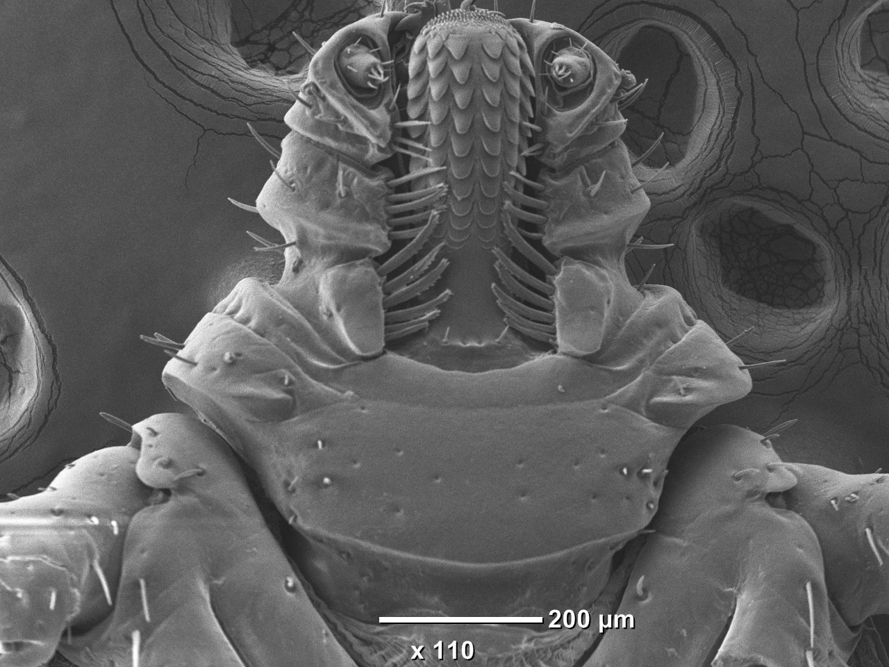
Below are the photos submitted for the "Best microscipic photo of a tick" category. All photos are copyrighted by their authors. The photos are listed in the order they were received.
MI1: Ladislav Simo - Amblyomma variegatum - Peptidergic innervation of hard tick salivary gland:

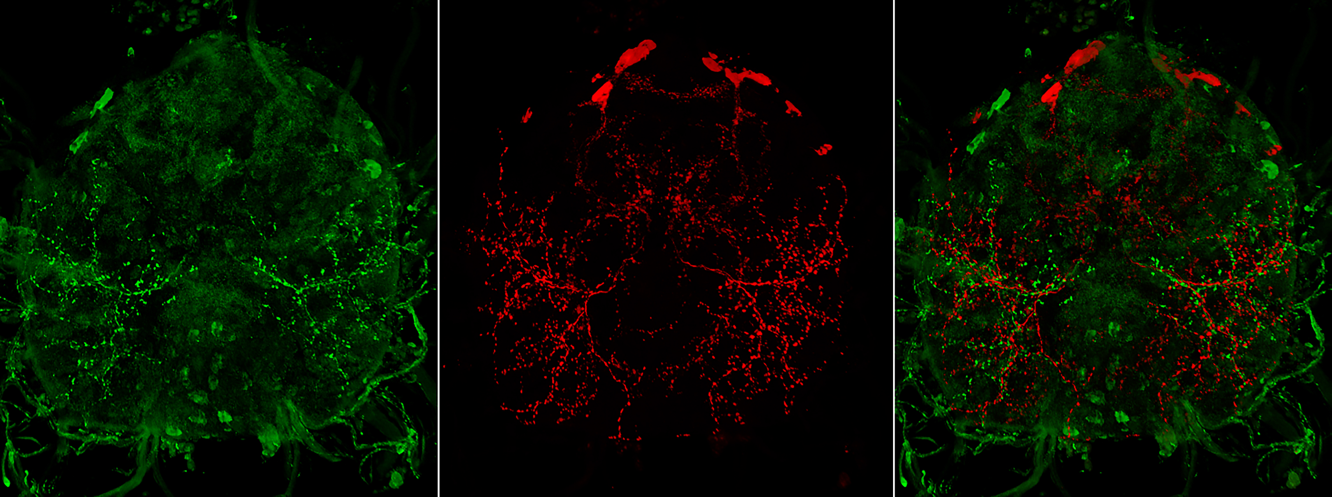
MI2: Ladislav Simo - Ixodes ricinus - Arborisation of neurosecretory axons on the surface of Ixodes synganglion:
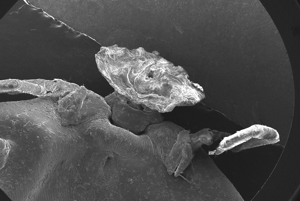
MI3: Jose de la Fuente - Rhipicephalus sanguineus - Cement cone:
MI4: Agina Onyinye - Theileria orientalis - Thin blood smear of Theileria orientalis infected crossbred Kedah-Kelantan x Brahman cattle:

MI5: Annetta Zintl - Ixodes ricinus - Beauty in the eye of the beholder.. or the lens of the confocal microscope:
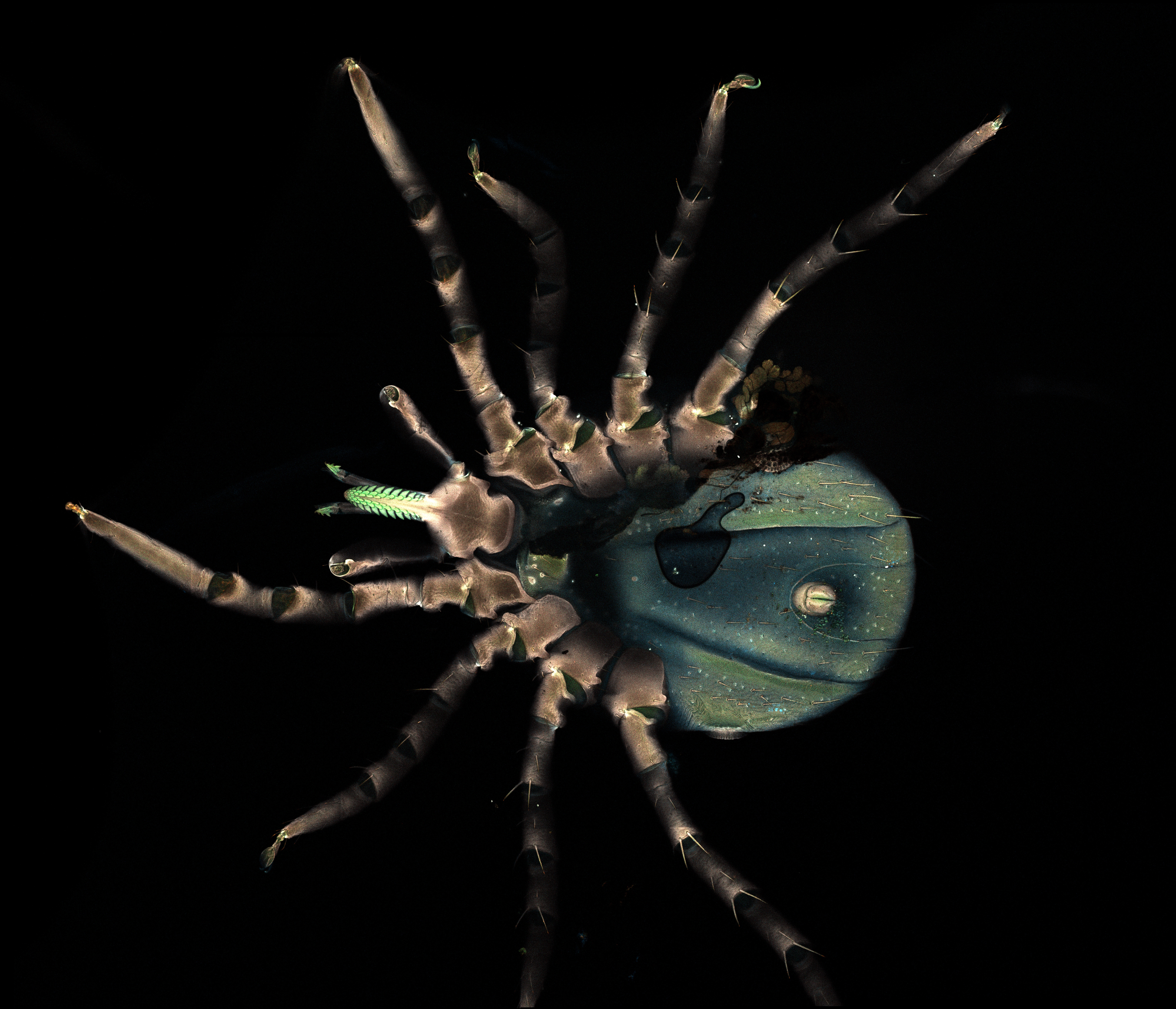
MI6: Alina Rodriguez Mallon - Amblyomma mixtum - Teeth, male:

MI7: Alina Rodriguez Mallon - Amblyomma mixtum - Spiracle, female:

MI8: Alina Rodriguez Mallon - Rhipicephalus microplus - Anus, female:

MI9: Alina Rodriguez Mallon - Rhipicephalus microplus - Haller organ, male:

MI10: Alina Rodriguez Mallon - Amblyomma mixtum - Palp, female:

MI11: Makwarela Tsireledzo Goodwill - Amblyomma hebraeum -dorsal side (left), ventral side (right):
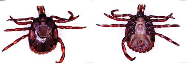
MI12: Makwarela Tsireledzo Goodwill - Hyalomma truncatum - dorsal side (left), ventral side (right):

MI13: Makwarela Tsireledzo Goodwill - Hyalomma rufipes - dorsal side (left), ventral side (right):

MI14: Makwarela Tsireledzo Goodwill - Rhipicephalus simus - dorsal side (left), ventral side (right):
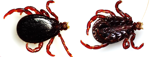
MI15: Makwarela Tsireledzo Goodwill - Rhipicephalus appendiculatus - dorsal side (left), ventral side (right):
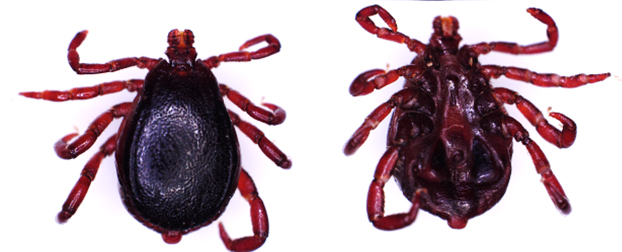
MI16: Ala Tabor - Haemaphysalis bancrofti - Microscopic picture of unfed adult female with larvae found attached to it (not visible by naked eye) and also a nymph:
 and also a nymph.jpg)
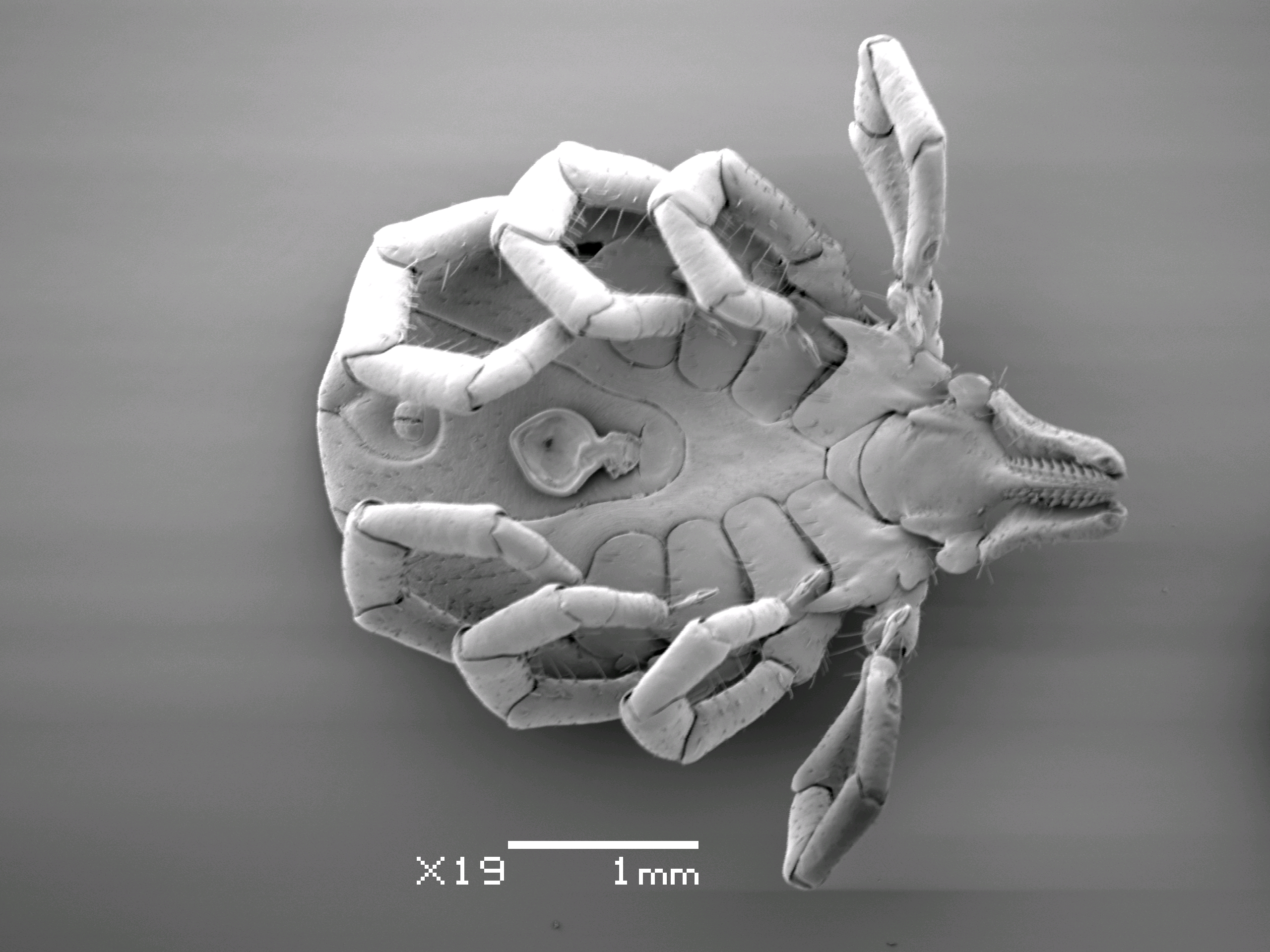
MI17: Ala Tabor - Ixodes holocyclus - SEM of female tick directly after mating – note the deposit:
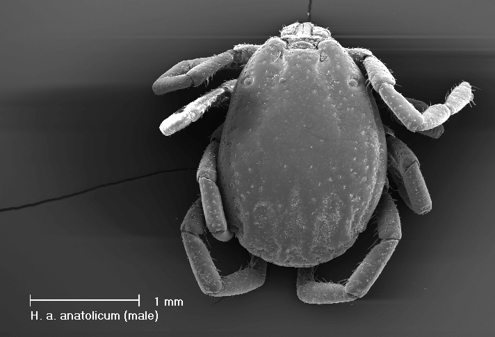
MI18: Nadira Akhtar - Hyalomma anatolicum anatolicum - SEM image (1):
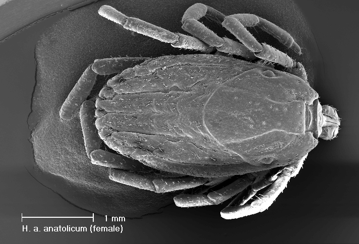
MI19: Nadira Akhtar - Hyalomma anatolicum anatolicum - SEM image (2):
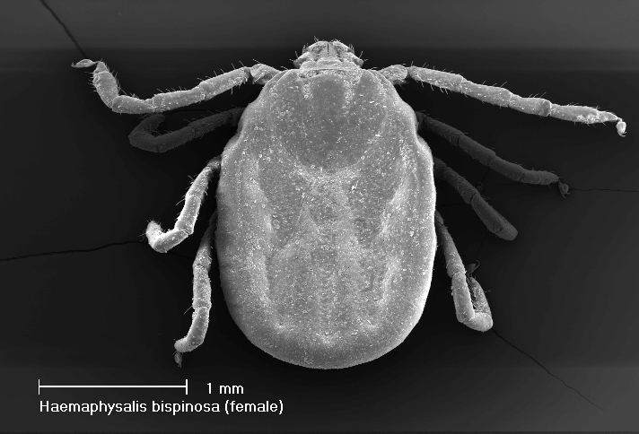
MI20: Nadira Akhtar - Haemaphysalis bispinosa - SEM image (1):
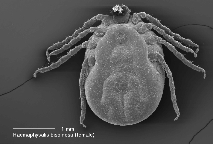
MI22: Nadira Akhtar - Haemaphysalis bispinosa - SEM image (2):
MI23: Sarah Bonnet & Thierry Blisnick - Ixodes ricinus - In a bad mood today:
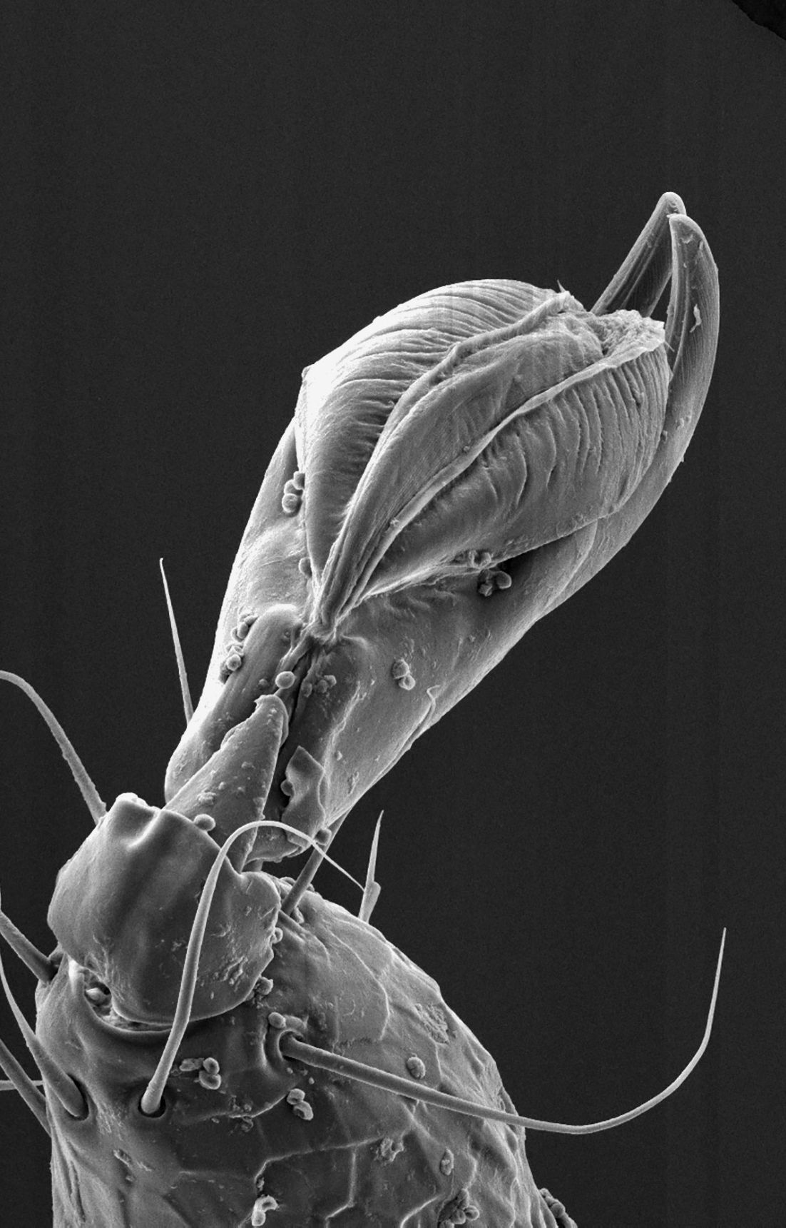
MI24: Sarah Bonnet & Thierry Blisnick - Ixodes ricinus - Tick's lace:
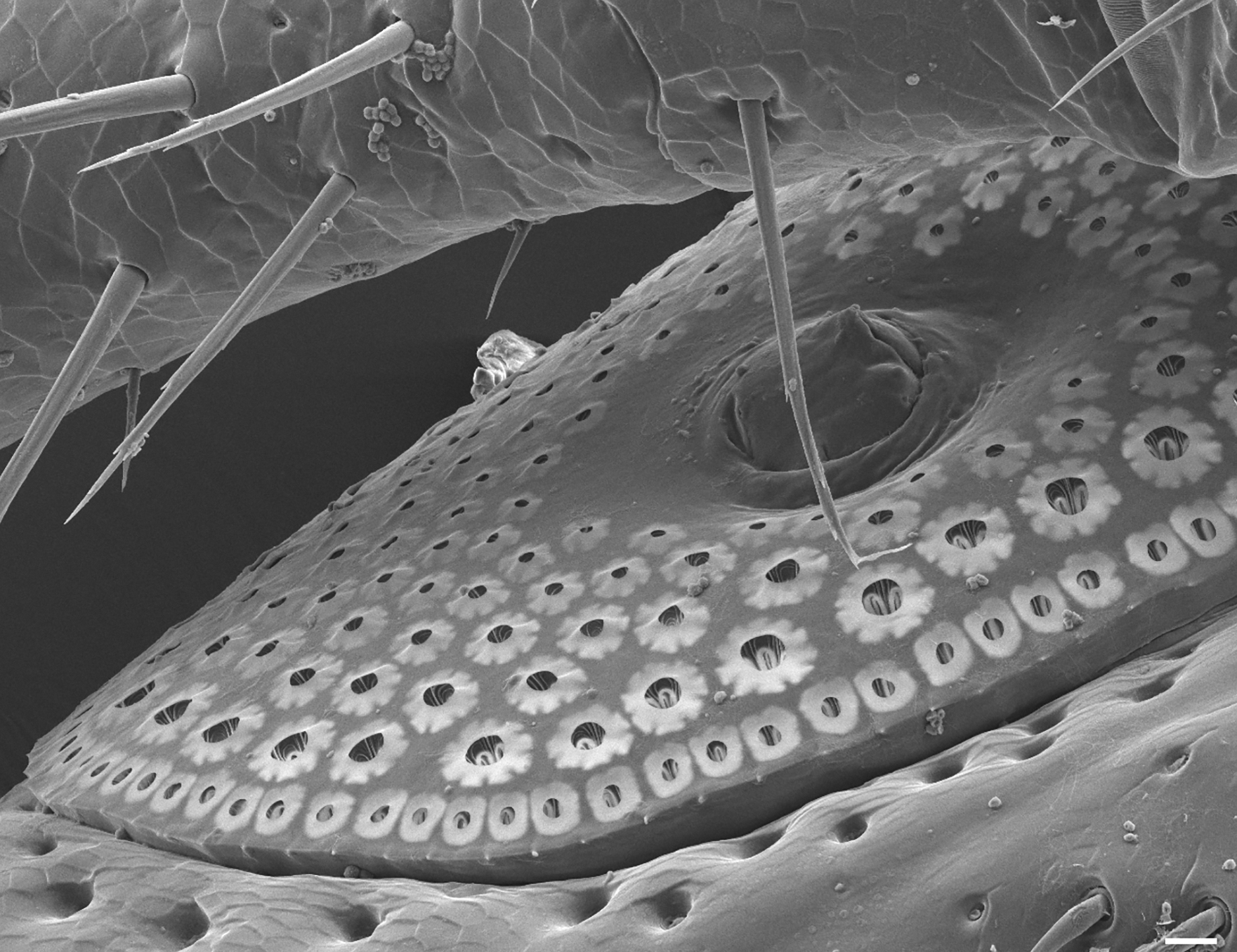
MI25: Sarah Bonnet & Thierry Blisnick - Ixodes ricinus - I know where you are:
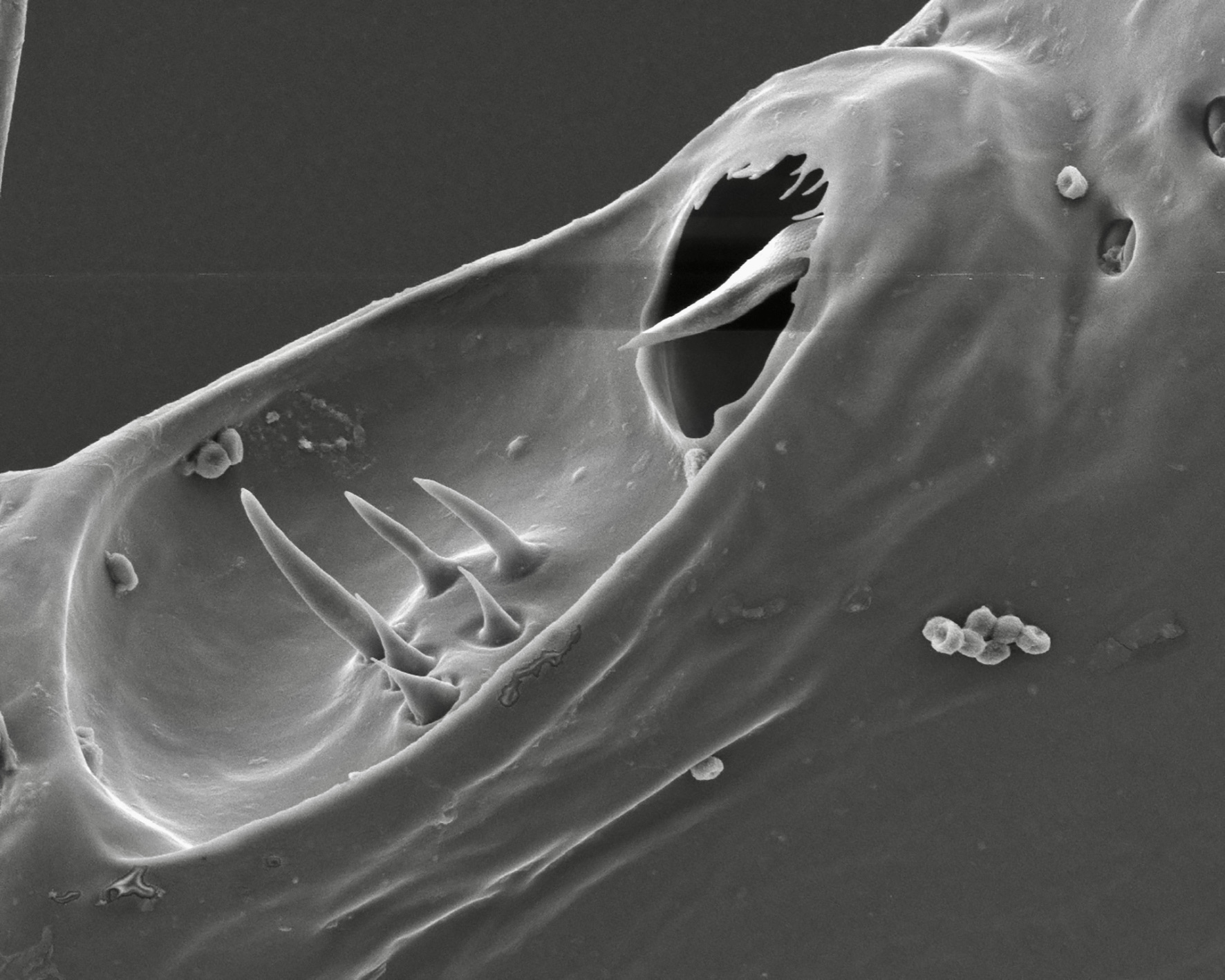
MI26: Gerhard Dobler - Ixodes rasus - female, In action:
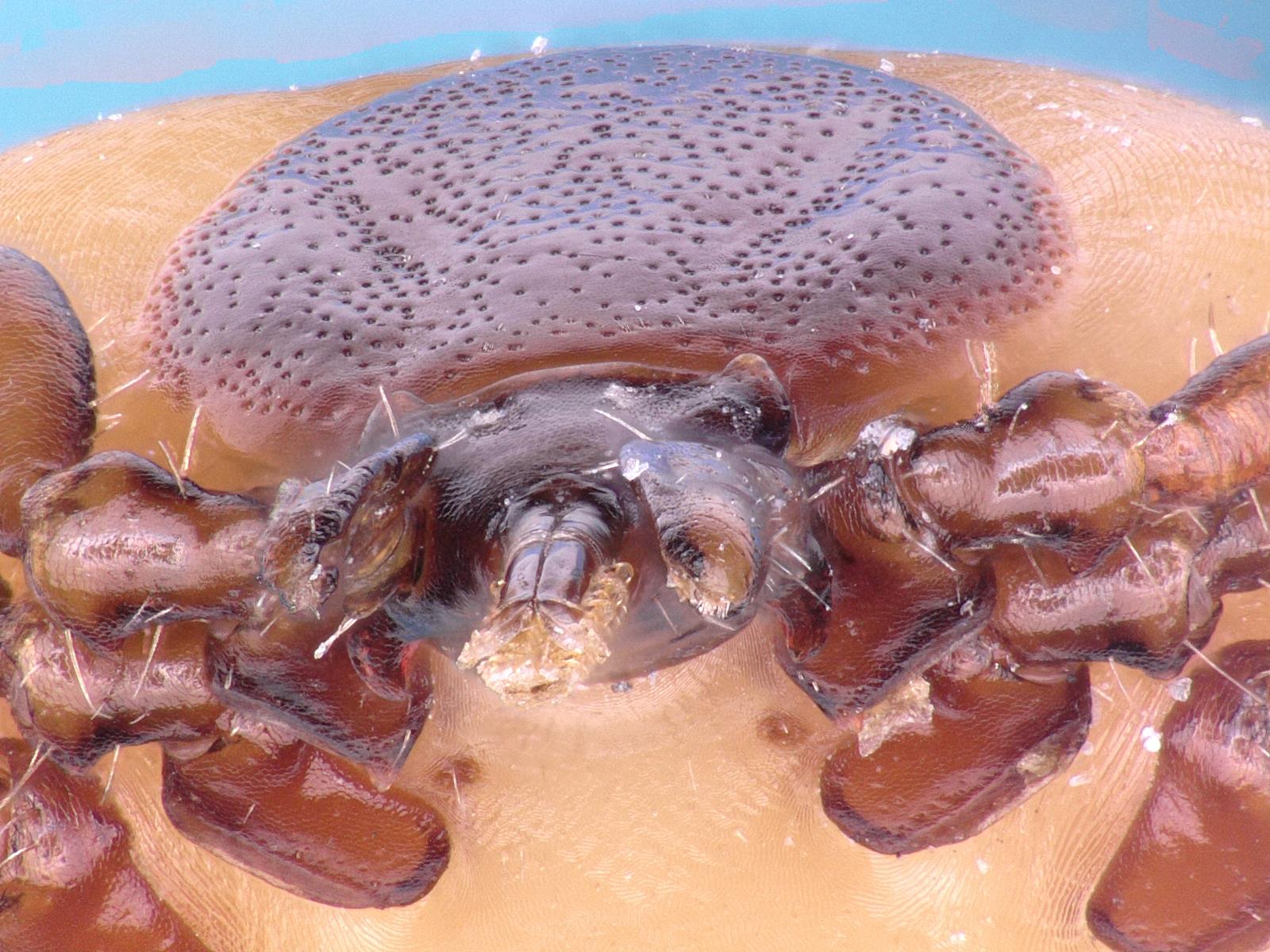
MI27: Gerhard Dobler - Ixodes ricinus - Tick in ice:

MI28: Lidia Chitimia-Dobler - Cornupalpatum burmanicum - female, tarsus I, hang a dinosaur feather, Never give up:

MI29: Lidia Chitimia-Dobler - Amblyomma tonelliae - female, Tick from Argentina:

MI30: Lidia Chitimia-Dobler - Haemaphysalis leachi - male, Beauty:
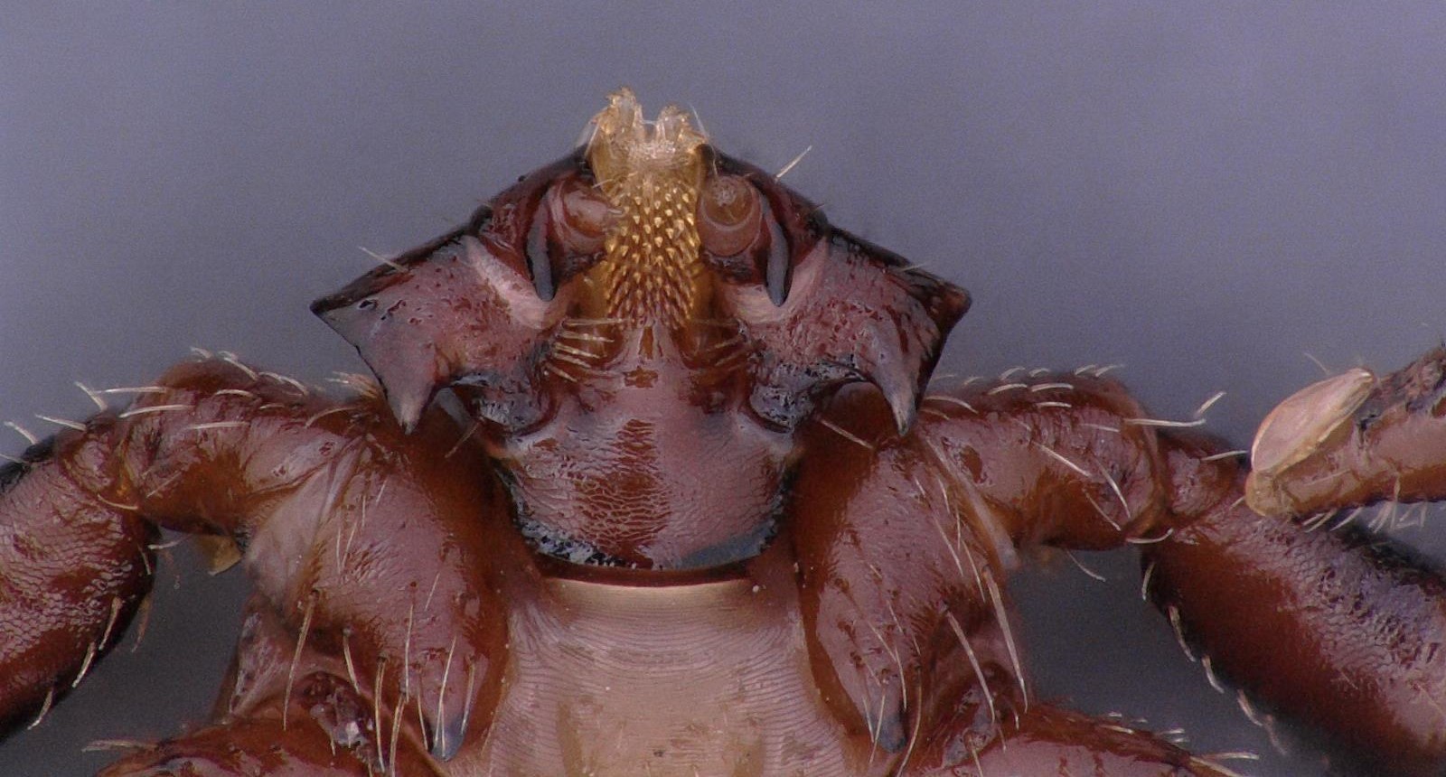
MI31: Lidia Chitimia-Dobler - Nosomma monstrosum - male, Not a real monster:

MI32: Lidia Chitimia-Dobler - Rhipicephalus pulchellus - female, Zebra tick:
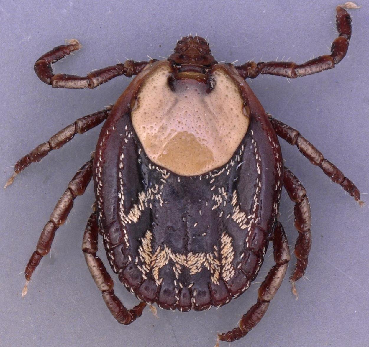
MI33: Lidia Chitimia-Dobler - Ixodes frontalis - female, Halloween tick:
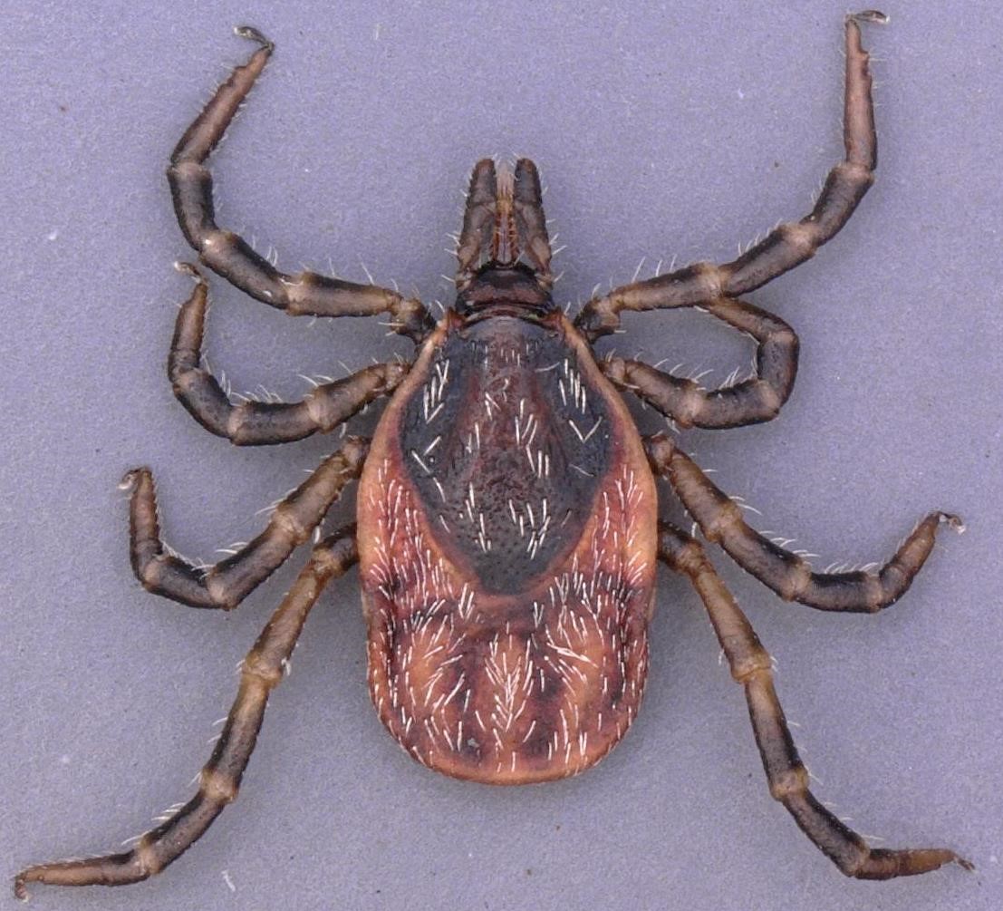
MI34: Ning Caina - Ixodes ricinus - Axon terminals (green) in acini type2 of hard tick salivary gland:
 in acini type2 of hard tick salivary gland.jpg)
MI35: Esther Kanduma - Rhipicephalus apendiculatus - SEM image showing ventral view of mouth parts